
The Adrenal Glands Explained: Cortex, Medulla, and the Stress Hormones
Endocrine biology · Glands behind the axes
The adrenal glands are two small, triangular organs that sit one on top of each kidney. Despite their size, they are really two glands in one: an outer cortex that manufactures steroid hormones, and an inner medulla that pumps out the fast “fight-or-flight” catecholamines. Understanding the adrenal glands ties together stress, salt balance, metabolism, and several of the peptides researchers discuss.

Two glands in one, with two separate origins
The adrenal (or suprarenal) glands have a striking split personality that even shows up in how they form. The outer cortex develops from mesoderm and makes steroid hormones from cholesterol. The inner medulla develops from neural crest — the same tissue that builds parts of the nervous system — and behaves like a specialized piece of the sympathetic nervous system. So a single gland houses a slow, gene-level chemical system and a near-instant neural one, side by side.
The cortex: three zones, three kinds of steroid
The cortex is organized into three layers, easy to remember with the mnemonic “GFR — salt, sugar, sex”:
- Zona glomerulosa (outer) makes mineralocorticoids, chiefly aldosterone — the “salt” hormone that helps manage sodium and blood pressure.
- Zona fasciculata (middle) makes glucocorticoids, chiefly cortisol — the “sugar”/stress hormone.
- Zona reticularis (inner) makes adrenal androgens such as DHEA and DHEA-S — the “sex” steroids.
All three are steroids built from cholesterol. The committed first step is the same everywhere: a transport protein (StAR) shuttles cholesterol into the mitochondria, where the enzyme CYP11A1 converts it to pregnenolone — the rate-limiting step of steroid production. What makes each zone different is the specific set of downstream enzymes it carries, which routes that shared precursor toward aldosterone, cortisol, or androgens.
The medulla: the body’s instant response
At the core, chromaffin cells of the medulla make the catecholamines epinephrine (adrenaline) and norepinephrine (noradrenaline), built from the amino acid tyrosine. The medulla is wired directly into the sympathetic nervous system: preganglionic nerve fibers synapse straight onto the chromaffin cells, which then dump their hormones into the bloodstream within seconds — the classic fight-or-flight surge.
There is an elegant link between the two halves here. The enzyme PNMT, which converts norepinephrine into epinephrine, is found almost exclusively in the adrenal medulla, and it is switched on by cortisol. Because cortisol-rich blood from the cortex drains inward through the medulla, the inner gland is bathed in the very hormone it needs to keep making adrenaline — the outer gland quietly enabling the inner one.
Three hormones, three different controllers
One of the most useful things to know about the adrenal glands is that their three main outputs answer to three completely different control systems.

- Cortisol is run by the HPA axis: the hypothalamus releases CRH, the pituitary releases ACTH, and ACTH binds the MC2R receptor on the cortex to drive cortisol. Circulating cortisol then feeds back to switch CRH and ACTH off.
- Aldosterone is controlled mainly by the renin-angiotensin-aldosterone system and by blood potassium — not primarily by ACTH. Low blood flow to the kidney triggers renin, leading to angiotensin II, which (along with high potassium) stimulates the glomerulosa.
- Catecholamines are controlled by the sympathetic nerves that fire directly onto the medulla.
What cortisol actually does
Cortisol is far more than a “stress hormone.” It raises blood glucose by promoting gluconeogenesis and opposing insulin, mobilizes fat, dampens the immune system (the basis of glucocorticoid anti-inflammatory drugs), and has permissive cardiovascular effects — it must be present for catecholamines to fully support blood pressure. Its release follows a daily rhythm, peaking in the early morning and reaching its low point around midnight.
When the adrenal glands go wrong
A few named conditions help define the gland’s range (for background only, not diagnosis): Cushing syndrome is excess cortisol; Addison disease is primary adrenal insufficiency, where the gland cannot make enough cortisol; and a pheochromocytoma is a catecholamine-secreting tumor of the medulla that causes surges of adrenaline and noradrenaline.
Why this matters for peptide research: the melanocortin map
ACTH is a melanocortin, cut from the same precursor protein (POMC) as the MSH peptides. But ACTH acts on its own receptor, MC2R, which is essentially adrenal-specific and drives cortisol. The MSH-type peptides act on the other melanocortin receptors — MC1R, MC3R, MC4R, and MC5R — which handle pigment, appetite/energy, and other roles.
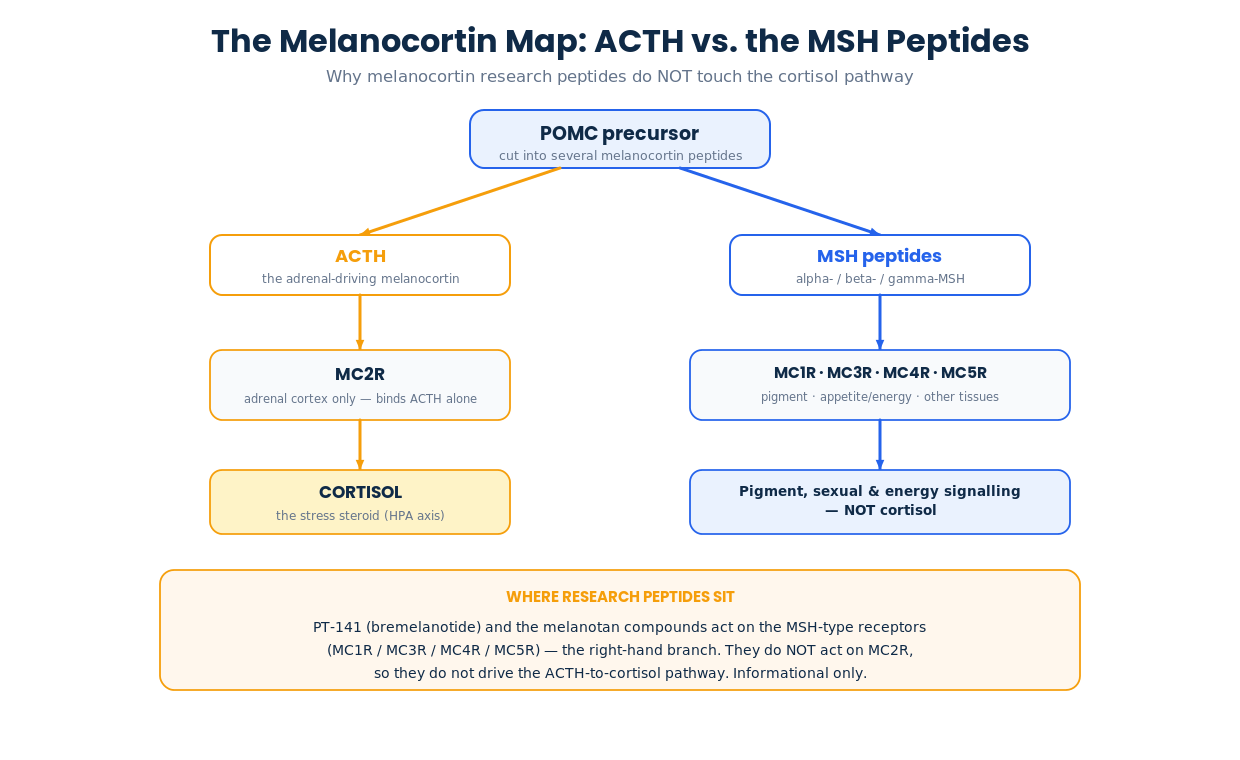
This is why melanocortin research peptides such as PT-141 (bremelanotide) and the melanotan compounds, which act on the MSH-type receptors, do not engage the ACTH-to-cortisol pathway. It also explains why exogenous steroids suppress the HPA axis: extra glucocorticoid feeds back on the hypothalamus and pituitary, quieting CRH and ACTH. All of this is informational background, not medical or dosing advice.
Frequently asked questions
What do the adrenal glands do in one sentence?
They make steroid hormones (aldosterone, cortisol, and androgens) in the cortex and fast-acting catecholamines (adrenaline and noradrenaline) in the medulla.
Are adrenal hormones peptides?
No — the cortex makes steroids from cholesterol and the medulla makes catecholamines from tyrosine. The peptide connection is upstream: ACTH, the pituitary peptide that tells the cortex to make cortisol.
Why is cortisol called a stress hormone?
Because the HPA axis ramps up cortisol during stress, and cortisol then mobilizes energy, modulates the immune system, and supports blood pressure to help the body cope.
Do melanocortin peptides affect cortisol?
Not through the adrenal receptor. ACTH uses MC2R to drive cortisol, while MSH-type melanocortin peptides act on MC1R/MC3R/MC4R/MC5R. This is background biology, not advice.
References
- Dutt M, Wehrle CJ, Jialal I. Physiology, Adrenal Gland. StatPearls, NCBI Bookshelf.
- Megha R, Wehrle CJ, Kashyap S, Leslie SW. Anatomy, Abdomen and Pelvis: Adrenal Glands (Suprarenal Glands). StatPearls, NCBI Bookshelf.
- Nicolaides NC, et al. Adrenal Cortex: Development, Anatomy, Histology and Physiology. Endotext, NCBI Bookshelf.
- Clark AJL, et al. ACTH Action on the Adrenals. Endotext, NCBI Bookshelf.
- Glucocorticoid Therapy and Adrenal Suppression. Endotext, NCBI Bookshelf.
- Molecular machinery of catecholamine biosynthesis (PNMT regulation by glucocorticoids). PMC.
Informational and educational only — not medical advice. Consult a qualified healthcare professional. Intended for adults 21+.