
Peptide Secondary Structure: Alpha Helix, Beta Sheet and Turns
Peptide Science
Peptide secondary structure is the local shape a short amino acid chain folds into — mainly alpha helices and beta sheets — held together entirely by hydrogen bonds within its own backbone. It’s not a cosmetic detail: secondary structure affects how stable a peptide is, how well it resists being broken down, and how tightly it can bind a target. Here’s what the structural biology literature actually documents about how these shapes form and how researchers study them.
Primary, secondary, and tertiary: three levels of structure
A peptide’s primary structure is just its amino acid sequence — the order of building blocks, read like letters in a word. Secondary structure is the first level of 3-D shape: local, repeating patterns the backbone folds into because of hydrogen bonds between its own atoms, independent of what the side chains are doing. Tertiary structure is the fuller 3-D fold of an entire chain, secondary elements packed together into a working shape. This article is about that middle level — the two dominant local patterns, the alpha helix and the beta sheet, plus the turns that connect them.
The alpha helix: a coil held together by its own backbone
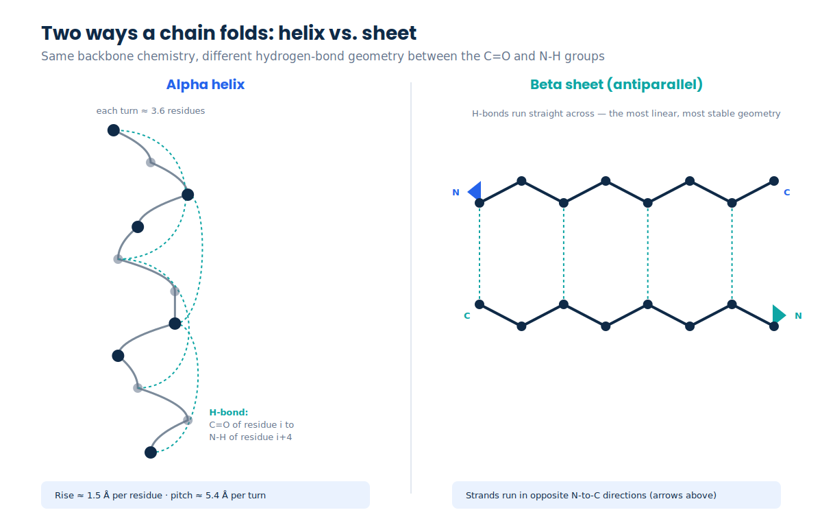
An alpha helix forms when a stretch of backbone coils into a right-handed spiral, stabilized entirely by hydrogen bonds within the chain itself: the carbonyl oxygen (C=O) of one residue bonds to the amide hydrogen (N-H) of the residue four positions further along — an “i to i+4” pattern. That single repeating bond, over and over, is what holds the coil together. Geometrically, a helix turns about every 3.6 residues, rising roughly 1.5 Å per residue (a full turn’s pitch is about 5.4 Å). Not every amino acid coils equally well: alanine, leucine and arginine are strong helix-formers, while proline — whose ring structure blocks the needed N-H hydrogen bond and kinks the backbone — is the strongest helix-breaker, and glycine’s extra flexibility also tends to destabilize a helix.
The beta sheet: strands held together side by side
A beta sheet forms differently: instead of one strand coiling onto itself, two or more extended strands of backbone line up side by side, hydrogen-bonding to each other rather than within a single strand. When neighboring strands run in opposite N-to-C directions, the arrangement is antiparallel, and its hydrogen bonds sit nearly straight across — the most linear, generally most stable geometry. When strands run the same direction, the sheet is parallel, with a slightly more bent, less optimal hydrogen-bond angle. Real strands aren’t perfectly flat — they twist slightly, which is part of why sheets keep their pleated look and don’t splay apart.
Turns: how a chain reverses direction
Helices and sheets are usually connected by short turns rather than running directly into each other. A beta turn is a compact, 4-residue motif stabilized by a single hydrogen bond between the carbonyl of residue i and the amide hydrogen of residue i+3 — a tighter loop than the helix’s i-to-i+4 pattern. Turns are almost always on the surface of a folded structure, and a beta-hairpin (two antiparallel strands connected by one tight turn) is one of the most common small motifs in known protein and peptide structures.
Why secondary structure matters for stability
A short, free peptide in solution is often more floppy and dynamic than a folded domain inside a large protein — there’s less surrounding structure to hold any one shape in place. That’s part of why “stapled peptides” exist as a deliberate engineering approach: chemists install a hydrocarbon “staple,” typically bridging residues one or two helical turns apart, that locks a short chain into a stable alpha-helical shape. Beyond just holding the shape, a properly stapled helix is measurably more resistant to protease degradation and can bind its target with higher affinity — which is exactly why stapled peptides are being pursued as drug candidates against targets that are otherwise hard to reach with a conventional peptide or small molecule.
When folding goes wrong: aggregation and amyloid
Beta-sheet structure specifically, in a particular arrangement called a parallel, in-register cross-beta structure, is the defining architecture of amyloid fibrils — the aggregates associated with conditions like Alzheimer’s disease. X-ray microcrystallography of amyloid-forming peptide segments has shown the fibril “spine” is built from a pair of beta sheets forming a dry, tightly interdigitated interface researchers call a steric zipper, with extensive backbone hydrogen bonding keeping strands locked in register — which is part of why amyloid fibrils are so physically and chemically stable once formed. Solid-state NMR studies of amyloid-beta specifically show ordered, in-register parallel beta strands connected by a flexible loop region. Importantly, the research literature is explicit that “considerable uncertainty currently exists” about exactly which aggregated forms are most responsible for cellular toxicity — the structural transition toward cross-beta sheet is well documented; the precise toxicity mechanism is still an active research question, not a settled one.
How researchers actually determine secondary structure
No single method covers every case, which is why structural biology leans on several complementary techniques. Circular dichroism (CD) spectroscopy is fast and solution-based, estimating the overall percentage of helix, sheet and disordered content from how a peptide’s far-UV light spectrum is shaped — useful for a quick read, but with no atomic-level detail. X-ray crystallography, the source of most structures in the Protein Data Bank, diffracts X-rays off an ordered crystal to build a detailed electron-density map, though it requires the protein to form a crystal in the first place and can miss flexible regions entirely. NMR spectroscopy studies a peptide in solution rather than locked in a crystal, making it the go-to method for small, flexible chains, while cryo-EM images thousands of frozen individual particles and reconstructs a 3-D density map computationally — a technique that now rivals crystallography for many targets, especially large assemblies.

Once a 3-D structure exists — solved experimentally — a standard algorithm called DSSP (Define Secondary Structure of Proteins) assigns each residue a label (helix, strand, turn, and so on) purely from its hydrogen-bond pattern. That’s structure assignment, working backward from a known shape. Structure prediction is a different problem: AlphaFold, evaluated at the CASP14 competition, predicted full atomic coordinates directly from sequence with a median backbone accuracy around 0.96 Å — roughly three times more accurate than the next-best prior method — even for sequences with no close structural template. A predicted structure’s secondary-structure content follows from those coordinates the same way DSSP derives it from an experimental one.
Cautions and considerations
This article describes established structural biology for research and educational purposes. It is not medical advice, and no part of it is a protocol for handling, dosing, or administering any compound. Specific structural claims about a named peptide (for example, exactly how helical a given hormone is in its receptor-bound state) should be checked against a primary structural paper before being treated as fact — several plausible-sounding numbers in this space could not be independently confirmed during research for this article and were deliberately left out.
Frequently asked questions
What’s the difference between primary, secondary, and tertiary structure?
Primary structure is the amino acid sequence itself. Secondary structure is the local folding pattern — mainly alpha helices and beta sheets — held together by hydrogen bonds within the backbone. Tertiary structure is the complete 3-D shape of one chain, built from secondary elements packed together.
Why does a peptide’s secondary structure matter for stability?
A defined shape, like a locked alpha helix, tends to resist enzymatic degradation better than a floppy, disordered chain — which is the whole rationale behind engineering approaches like peptide stapling. Structure also affects how tightly a peptide can bind its target.
How do researchers figure out a peptide’s secondary structure?
Mainly through circular dichroism, X-ray crystallography, NMR spectroscopy and cryo-EM — each with different trade-offs between speed, atomic detail, and the physical state the sample has to be in. Computational tools like DSSP and AlphaFold then assign or predict structure from those results (or from sequence alone, in AlphaFold’s case).
Can a short peptide form a stable secondary structure on its own?
Often not as stably as a folded domain inside a larger protein — short free peptides in solution tend to be more dynamic. That’s precisely why constrained-design approaches like hydrocarbon stapling exist: to lock a short sequence into a stable helix on purpose, rather than leaving it to fold (or not) on its own.
References
- “Alpha helix” — helix geometry, hydrogen-bond pattern, helix-forming/breaking residues. en.wikipedia.org/wiki/Alpha_helix
- “Beta sheet” — parallel vs antiparallel strand geometry and amyloid cross-beta structure. en.wikipedia.org/wiki/Beta_sheet
- Nelson R, Sawaya MR, Balbirnie M, et al. “Structure of the cross-beta spine of amyloid-like fibrils.” pmc.ncbi.nlm.nih.gov/articles/PMC1479801
- Tycko R. “Molecular Structure of Aggregated Amyloid-beta: Insights from Solid-State NMR.” pmc.ncbi.nlm.nih.gov/articles/PMC4968170
- Jumper J, Evans R, Pritzel A, et al. “Highly accurate protein structure prediction with AlphaFold.” Nature 596 (2021). nature.com/articles/s41586-021-03819-2
- “Methods for Determining Structure” — PDB-101, RCSB Protein Data Bank. pdb101.rcsb.org (methods guide)
- “All-Atom Model for Stabilization of Alpha-Helical Structure in Peptides by Hydrocarbon Staples.” pmc.ncbi.nlm.nih.gov/articles/PMC2735086
Informational only — not medical advice · 21+. This article describes established structural biology for research and educational purposes and is not a protocol for handling, dosing or administering any compound.