
Second Messengers Explained: How Cells Relay a Hormone Signal
Cell Signaling
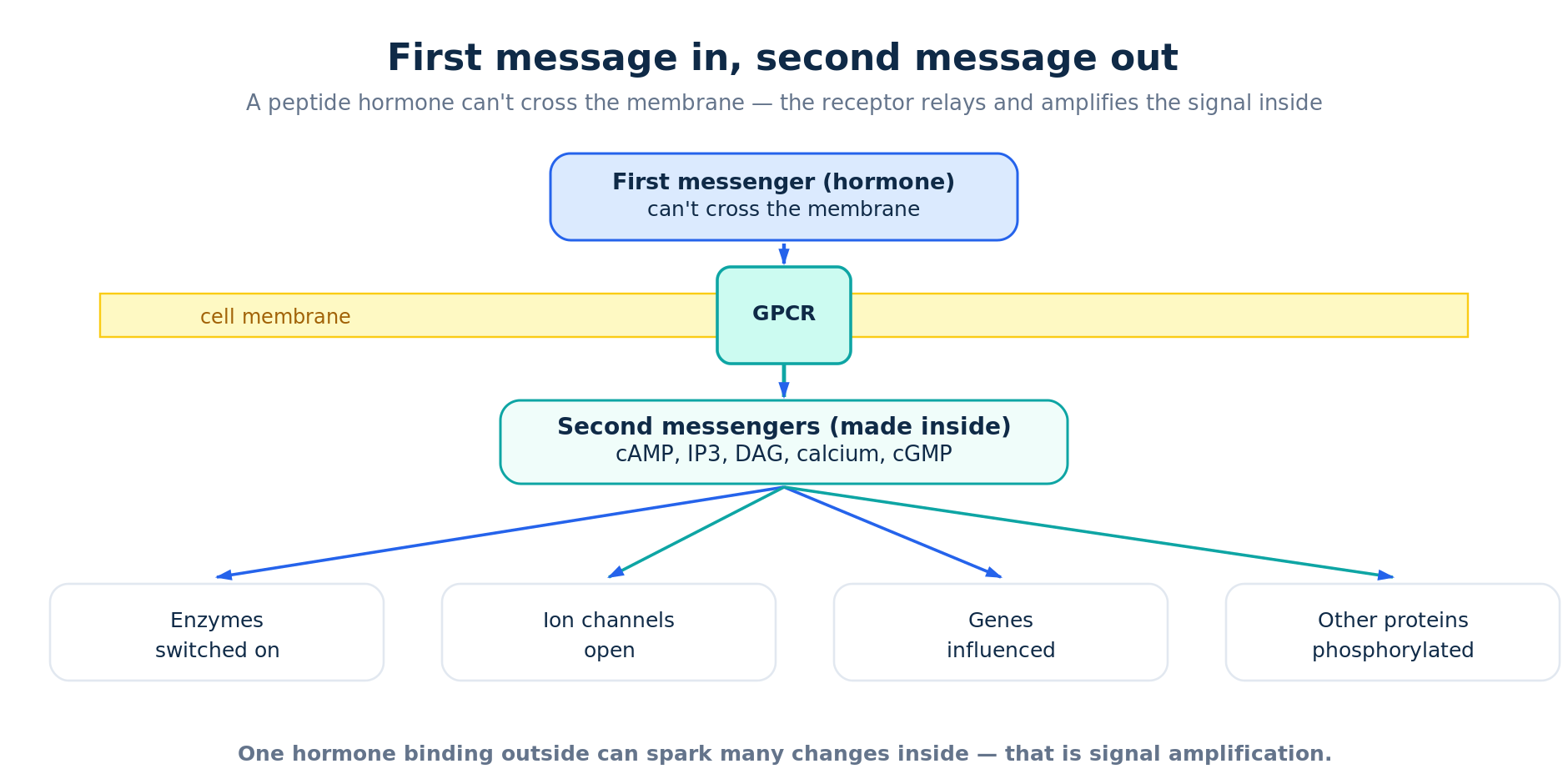
Second messengers are the small molecules a cell makes inside itself to carry a hormone’s message inward. A peptide hormone usually cannot cross the fatty cell membrane, so it binds a receptor on the surface — often a G-protein-coupled receptor — and that receptor triggers a burst of intracellular messengers such as cAMP, calcium and others. Those messengers relay and amplify the signal, then are quickly switched off. It is the reason peptide-hormone effects tend to be fast and reversible.
The membrane problem: first and second messengers
Biologists call the hormone the “first messenger” and the intracellular relay molecule the “second messenger.” The idea traces back to Earl Sutherland, who discovered cyclic AMP (cAMP) while working out how adrenaline and glucagon make the liver release glucose — work recognised with the 1971 Nobel Prize. The key insight: the hormone itself often never enters the cell. The receptor converts the outside message into an inside one.
The cAMP pathway
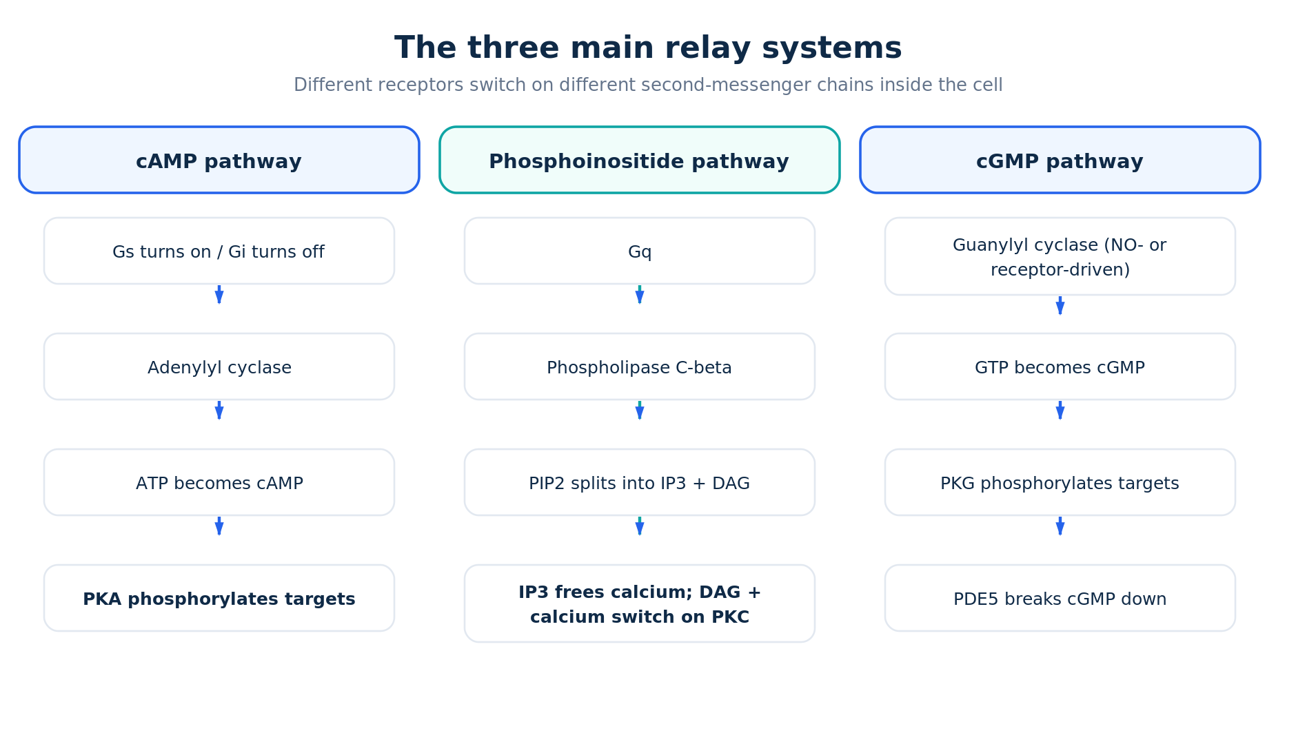
The best-studied system runs on cAMP. When a hormone binds a receptor coupled to a stimulatory G protein (Gs), it activates the membrane enzyme adenylyl cyclase, which converts ATP into cAMP. A receptor coupled to an inhibitory G protein (Gi) does the opposite and turns the enzyme down — so the same messenger can be raised or lowered depending on the receptor. cAMP’s main job is to switch on protein kinase A (PKA), which then phosphorylates target proteins and can even reach the nucleus to influence gene activity through the transcription factor CREB. Hormones such as glucagon and ACTH act largely through this Gs–cAMP route.
The phosphoinositide pathway: IP3 and DAG
A second major system is triggered by receptors coupled to the Gq protein, which activates phospholipase C-beta. That enzyme splits a membrane lipid called PIP2 into two messengers at once: IP3 and DAG. IP3 is water-soluble and diffuses to the endoplasmic reticulum, where it opens channels that release stored calcium into the cell. DAG stays in the membrane and, together with calcium, switches on protein kinase C (PKC). So a single receptor event produces three converging signals — IP3, DAG and a rise in calcium.
Calcium and cGMP
Calcium is itself a universal second messenger. Cells normally keep the amount in their cytoplasm very low, so even a small, controlled release from internal stores is a strong signal; much of calcium’s action is carried out through the protein calmodulin. A parallel cyclic nucleotide, cGMP, is made from GTP by guanylyl cyclase. One form of that enzyme is switched on by nitric oxide; another form is itself a surface receptor — for example the receptor for natriuretic peptides is a guanylyl cyclase, not a GPCR. cGMP acts mainly through protein kinase G. This pathway is also a well-known drug target: PDE5 inhibitors work by slowing the breakdown of cGMP.
Amplify, then switch off
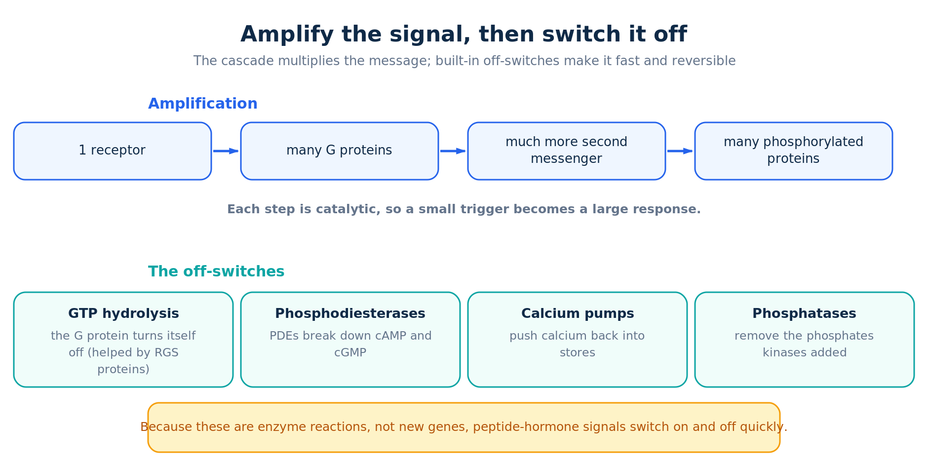
Two features make these systems powerful. First, they amplify: because each step is catalytic and branching, one activated receptor can switch on many G proteins, each enzyme makes many messenger molecules, and each kinase modifies many targets — so a faint outside signal becomes a large inside response. Second, they have built-in off-switches: the G protein turns itself off by hydrolysing GTP, phosphodiesterases (PDEs) destroy cAMP and cGMP, pumps return calcium to storage, and phosphatases strip the phosphate groups back off. Because these are enzyme reactions rather than changes in gene transcription, the whole response can be switched on and off quickly.
Fast versus slow: this is the mirror image of steroid and thyroid hormones, which cross the membrane and act directly on genes through nuclear receptors — a slower, longer-lasting route. Same body, two very different signalling strategies.
Why it matters
Second messengers explain a great deal of pharmacology. Caffeine and related methylxanthines block PDEs; PDE5 inhibitors raise cGMP; beta-agonists and beta-blockers act on the Gs–cAMP system. Understanding which messenger a receptor uses also clarifies why two hormones acting on the same cell can either cooperate or oppose each other. For the peptides discussed across this site, the surface-receptor-plus-second-messenger model is usually the relevant one — and it stands in contrast to how the mature peptides themselves are built (prohormone processing).
Frequently asked questions
What is the difference between a first and second messenger?
The first messenger is the extracellular signal — usually the hormone. The second messenger is the small intracellular molecule (such as cAMP or calcium) the cell makes in response, which then spreads the signal inside.
What are the main second messengers?
The most important are cAMP, cGMP, calcium, and the pair IP3 and DAG produced from membrane lipids. Each is generated or released by its own receptor-driven pathway and acts on its own set of target proteins.
Why does the cell bother with second messengers instead of letting the hormone in?
Most peptide hormones are water-soluble and cannot cross the fatty membrane. Using a surface receptor plus internal messengers also lets the cell amplify a tiny signal and shut it off rapidly — something direct entry could not do as cleanly.
How is the signal turned off?
Several ways at once: the G protein hydrolyses its GTP, phosphodiesterases break down cAMP and cGMP, calcium is pumped back into storage, and phosphatases reverse the kinases’ work.
References
1. Molecular Biology of the Cell — Signaling through G-Protein-Linked Cell-Surface Receptors (NCBI Bookshelf). ncbi.nlm.nih.gov/books/NBK26912
2. Neuroscience (Purves) — Second Messengers (NCBI Bookshelf). ncbi.nlm.nih.gov/books/NBK10794
3. StatPearls — Biochemistry, cAMP (NCBI Bookshelf). ncbi.nlm.nih.gov/books/NBK535431
4. StatPearls — Biochemistry, Cyclic GMP (NCBI Bookshelf). ncbi.nlm.nih.gov/books/NBK542234
5. The Nobel Prize in Physiology or Medicine 1971 — Earl W. Sutherland Jr. (cAMP). nobelprize.org (1971)
6. The Nobel Prize in Physiology or Medicine 1994 — Gilman & Rodbell (G proteins). nobelprize.org (1994)
Informational only — not medical advice. For research and educational purposes; intended for audiences 21+. Always consult a qualified healthcare professional.