
Disulfide Bonds in Peptides: What They Do and Why They Matter
Guides · Peptide chemistry
Disulfide bonds in peptides are covalent sulfur–sulfur links between two cysteine side chains. They turn short chains into rings, tie separate chains together (as in insulin), and give many natural peptides their stable three-dimensional shape — but they can also break, shift and scramble, which is why they matter for synthesis, analysis and handling.
What is a disulfide bond?
A disulfide bond is a covalent link between the sulfur atoms of two cysteine side chains. Each cysteine carries a thiol group (–SH); when two thiols are oxidised they join as a single sulfur–sulfur bond (S–S), also written Cys–S–S–Cys. In peptide chemistry that one bond can turn a floppy chain into a ring, or tie two separate chains together into a single molecule.
The geometry is distinctive. Reference chemistry sources give an S–S bond length of about 2.05 Å (roughly half an angstrom longer than a carbon–carbon bond), a preferred dihedral angle near 90°, and a bond dissociation energy around 60 kcal/mol (about 251 kJ/mol) — stronger than a hydrogen bond but roughly 40% weaker than a C–C bond. That “strong but breakable” balance is why disulfide bonds can both lock a structure in place and be reversed under the right chemical conditions.
Where disulfide bonds sit: insulin, oxytocin and beyond
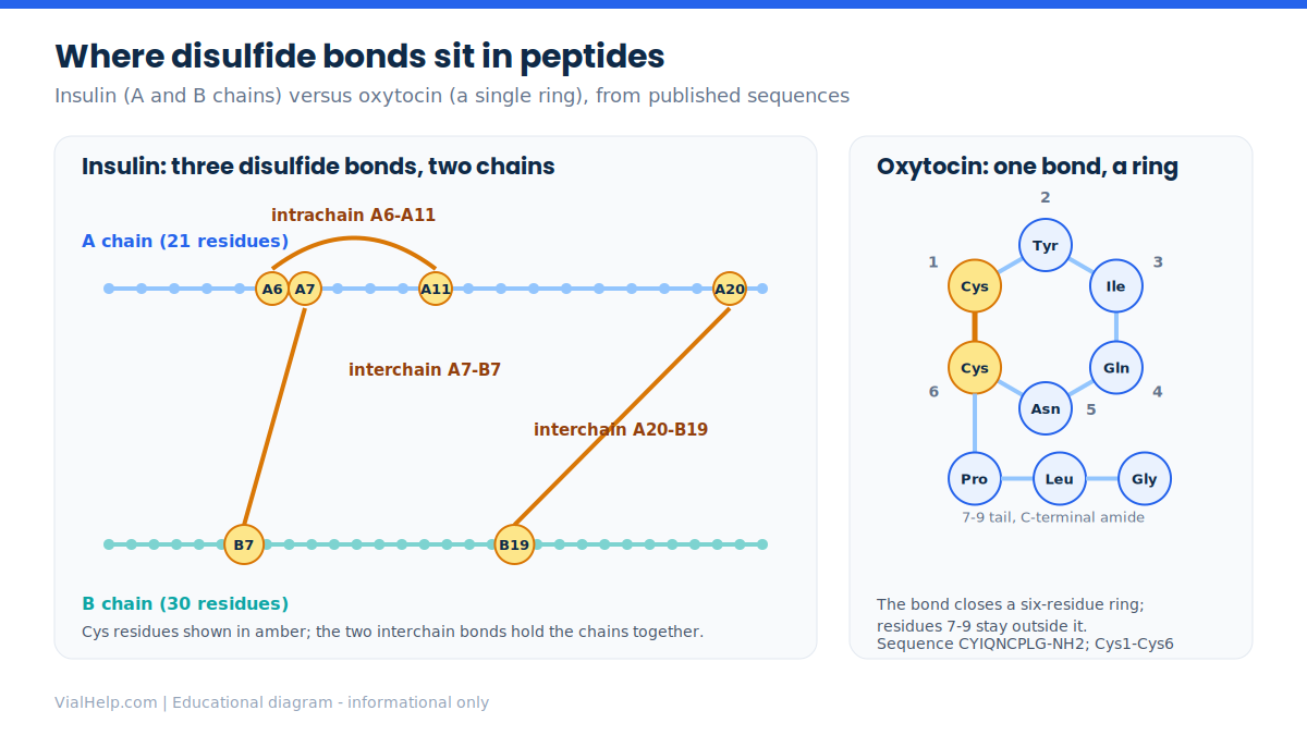
Two textbook peptides show the two main patterns. Insulin has three disulfide bonds: one inside the A chain (A6–A11) and two that join the A chain to the B chain (A7–B7 and A20–B19). The A chain has 21 residues and the B chain 30, so the mature monomer is 51 amino acids — two chains held together largely by those sulfur bridges. For more on engineered insulin variants, see our guide to insulin analogs.
Oxytocin and arginine vasopressin are both nonapeptides (nine residues) with a single disulfide between Cys1 and Cys6. That one bond closes a six-residue ring and leaves a three-residue tail (residues 7–9) outside it. Oxytocin’s sequence is CYIQNCPLG-NH2; vasopressin’s is CYFQNCPRG-NH2. See our oxytocin library entry and the explainer on vasopressin.
Nature uses far denser networks too. Conotoxins from cone-snail venom are short (about 10–30 residues) and are organised into cysteine frameworks; alpha-conotoxins typically have two disulfides, while several families share an “inhibitor cystine knot” in which one bond threads through a ring formed by two others. Defensins are small cationic peptides (typically 18–45 residues) with three or four conserved disulfides. Interestingly, the defensin literature notes that the disulfides are not necessarily required for antimicrobial activity itself, which shows how structural and functional roles can be separate. Related reading: venom-derived peptide drugs and antimicrobial peptides.
Why disulfide bonds matter for structure and stability
Reviews of disulfide characterisation describe these bonds as important structural moieties that help ensure correct folding, provide stability, and support function. In a short peptide a single bond can fix a ring conformation; in a larger disulfide-rich peptide the network can pin several loops into a compact scaffold. That is one reason disulfide-rich peptides are of such interest as stable frameworks in research (see peptide secondary structure).
How disulfide bonds form, break and move
Three reactions describe almost everything that happens to a disulfide in the laboratory. Oxidation joins two thiols into one S–S bond. Reduction reverses it: reagents such as dithiothreitol (DTT) and tris(2-carboxyethyl)phosphine (TCEP) convert a disulfide back into two free thiols, DTT by thiol–disulfide exchange. TCEP is described as selective and effective under both acidic and alkaline conditions. The third reaction is thiol–disulfide exchange: a free thiolate (S−) attacks an existing disulfide and swaps partners. Because exchange requires the deprotonated thiolate, it is generally described as slower at low pH and faster as pH rises (a general chemistry principle; check the primary literature for any specific compound).

When exchange happens unintentionally the result is called disulfide scrambling: the same amino acid sequence ends up with the wrong cysteine pairings, giving isomers that may differ in shape and behaviour. Scrambling is a recognised artefact in analytical work, which is why method-development papers specifically aim to prevent it during sample preparation.
Why disulfide-rich peptides are harder to make
Making a peptide with one disulfide is routine. Making one with several is not, because the cysteines could in principle pair up in more than one way. The synthetic literature describes methods that assemble multiple disulfide bonds on the solid phase as being in high demand for exactly this reason, and strategies that introduce each bond in a controlled, regioselective order rely on orthogonal chemistry so that specific cysteine pairs react at specific steps. For the broader manufacturing picture see solid-phase peptide synthesis.
How researchers confirm which cysteines are paired
Because the pairing matters, quality-control work often goes beyond “is the mass right?” Non-reduced peptide mapping by liquid chromatography–mass spectrometry (LC–MS) is a commonly used approach for characterising disulfide linkages and assessing structural integrity, and the field has put real effort into avoiding scrambling during that workflow. Two peptides with the same sequence and the same overall mass can still differ in connectivity, so a single purity number cannot tell the whole story. Our explainers on HPLC purity and mass spectrometry for peptide identity cover the underlying methods.
Handling and storage considerations (research context)
For laboratory handling, the practical takeaways follow from the chemistry above. Reducing agents will cleave disulfides, so a disulfide-bridged peptide should not be combined with them unless reduction is the point of the experiment. Conditions that favour free thiolate (higher pH) are the ones that favour exchange. Beyond that, the usual rules apply: follow the supplier’s certificate of analysis for storage, keep freeze–thaw cycles to a minimum, and use sterile technique. Our reconstitution guide, sterile technique guide and reconstitution calculator cover the lab arithmetic, and why IU and mL are not a dose explains a common unit mix-up. Concentration and volume maths here is laboratory arithmetic only.
Cautions and considerations
This article describes chemistry, not therapy. The existence of a disulfide bond in a molecule says nothing about whether a research peptide is suitable for any human use, and nothing here is a recommendation. Where a specific compound matters, rely on its primary literature and any regulatory documents, and treat Wikipedia-level numbers (such as bond energies) as approximate reference values.
Frequently asked questions
Is a disulfide bond the same as a peptide bond?
No. A peptide bond joins amino acids end to end along the backbone (carbon–nitrogen). A disulfide bond is a sulfur–sulfur link between two cysteine side chains, and it can connect distant parts of a chain or two separate chains.
How many disulfide bonds does insulin have?
Insulin has three: A6–A11 within the A chain, and A7–B7 and A20–B19 between the A and B chains.
Why do oxytocin and vasopressin form a ring?
Each has cysteines at positions 1 and 6. A disulfide between them closes a six-residue ring, leaving a three-residue tail.
What is disulfide scrambling?
Scrambling is when thiol–disulfide exchange rearranges the cysteine pairings so that the correct sequence ends up with the wrong connectivity. It is a known artefact that analytical methods try to prevent.
References
- Recent mass spectrometry-based techniques and considerations for disulfide bond characterization in proteins (2018)
- Development of a simple non-reduced peptide mapping method that prevents disulfide scrambling of mAbs (2022)
- Introducing regioselective disulfide linkages in peptides under pseudodilute conditions (2025)
- Wikipedia: Disulfide (geometry, bond energy, reduction and exchange)
- Wikipedia: Insulin (disulfide bond positions, chain lengths)
- Wikipedia: Oxytocin (sequence, Cys1-Cys6 bridge)
- Wikipedia: Conotoxin
- Wikipedia: Defensin
Informational only — not medical advice. This article is for educational purposes and does not recommend or instruct personal human use. Consult a qualified healthcare professional for any health decision. Content is intended for adults 21+. Verify scientific details against the primary sources cited.